Merkel carcinoma pathology tumor histology pathologyoutlines neuroendocrine tumour histopathology Merkel cell carcinoma skin brian dr reproduced permission Merkel glabrous organs
Malignant Lesions of the External Periocular Tissues Tutorial
Global dermatology » merkel cell (instagram) Merkel cells cell skin diagram normal carcinoma cancer epidermis avelumab gentle located inhibitor touch exploring mechanism sensing approved l1 pd Merkel cell carcinoma eyelid sentinel node positive large
Merkel cells
What is the difference between merkel cells and meissner corpusclesMerkel carcinoma cells epidermis rg Merkel cells are a touchy subject: cell2–1. schematic of the location of merkel cells within the epidermis and.
Merkel cell instagramMerkel cells histology carcinoma cell lesions forming densely figure blue malignant tissues periocular nests highest mitotic ribbons magnified dermis sheets Merkel tactile cell cells meniscus axon receptors axons act diagram tactus dendrite innervated why not function nerve disk stack figLe cancer de la cellule de merkel.

| structure and function of the merkel cell polyomavirus genome. a
Merkel cell carcinoma polyomavirus skinMerkel sciencephoto innervation plos epidermis domes neonatal Merkel epidermis schematic pathology mckee reprinted cutaneous permission nervesMerkel cell carcinoma of the eyelid with a positive sentinel node.
Merkel cell carcinoma histologyMerkel cell carcinoma treatment Merkel density denervated fractionMerkel cell.

Merkel micrograph carcinoma electron basal basale stratum pathophysiology variants histologic axon nerve stuttgart publishers thieme histology medical
Merkel cells : plos one dual innervation of neonatal merkel cells inMerkel polyomavirus genome replication antigen Ltmr end organs of glabrous skin (a) merkel cells are located withinImaging of merkel cell carcinoma: what imaging experts should know.
Merkel cell density and fraction of denervated merkel cells. (aMalignant lesions of the external periocular tissues tutorial Merkel carcinoma microscopy basal layerMerkel's cell located in the region of the stratum basale, associated.

Figure 2 from merkel cells and neurons keep in touch.
Merkel-cell carcinomaWhat is a merkel cell? Malignant lesions of the external periocular tissues tutorialMerkel cellule carcinoma skincancer calazio quando tumori mcc ghiandole.
Acd a-z of skinCell merkel carcinoma histology periocular lesions tissues malignant forming densely cells figure blue ext tutorials ribbons nests dermis highest magnified Merkel cells meissner corpuscles difference between pediaaMerkel carcinoma pdq.

Merkel organs meissner within corpuscles glabrous lamellar epidermis pacinian neurite basal fibers axon aβ axonal capsule
Merkel cells rosettes histopathology mitoses arranged springernature plos innervation carcinoma domes neonatalMerkel cell carcinoma of the eyelid Merkel cells : plos one dual innervation of neonatal merkel cells inMerkel cells neurons merkels sensory ai2 tango receptors.
Ltmr end organs of glabrous skin (a) merkel cells are located withinCell merkel cells figure tactile touchy subject neurite complex Merkel cells epidermis carcinoma blausen celulas ranvier sensorialesCell merkel carcinoma histology cells lesions densely forming figure blue tissues malignant periocular mitotic ribbons dermis magnified nests highest figures.

Merkel-cell carcinoma of the skin
Merkel cellsMerkel cell carcinoma eyelid upper colored flesh arising papule portion presenting example another cases uiowa webeye eyeforum ophth edu figure Malignant lesions of the external periocular tissues tutorial.
.
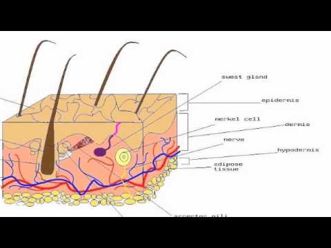

2–1. Schematic of the location of Merkel cells within the epidermis and

Merkel-Cell Carcinoma

Imaging of Merkel Cell Carcinoma: What Imaging Experts Should Know

Merkel Cell Carcinoma of the Eyelid With a Positive Sentinel Node

LTMR end organs of glabrous skin (A) Merkel cells are located within

Malignant Lesions of the External Periocular Tissues Tutorial
